Products
-

PARAMECIUM
Inquire nowThe model provides a detailed depiction of both external and internal features of the organism. One side highlights the oral groove,
-

PARAMECIUM MODEL
Inquire nowKey features identified include the pellicle, ectoplasm, endoplasm, trichocytes, peristome, vestibule, buccal cavity, gullet, cytopharynx, food vacuole, macronucleus, micronucleus,
-

Photon Energy Array
Inquire nowPhoton Energy Array
Seven LED’s of different wavelength mounted on an acrylic base bended at their edge to stand it.
-

Physics Lab
Inquire nowEducational Equipment
-

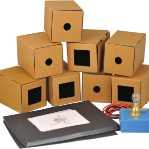
Pinhole Camera Kit
Inquire nowPinhole Camera Kit
Ideal for student experimentation in simple optics involving the basic pin hole camera. Other optical effects such as image inversion,
-

Pinhole Screen
Inquire nowCircular black metal screen about 75mm in diameter, with a central hole 0.6mm diameter approx.
-

PLACENTA MODEL
Inquire nowThe model shows the structure of placenta and the relation between placenta and umbilical card.
-

PLANT CELL ANATOMY STUDY MODEL
Inquire nowFeaturing key structures like the cell wall, plasma membrane, nucleus, chloroplasts, and more, it’s ideal for studying plant cell anatomy.
-

PLANT MITOSIS
Inquire nowPresenting a set of 10 models illustrating the complete process of karyokinesis and cytokinesis, from the metabolic cell to the formation of two daughter nuclei.
-

PLANT TISSUE CAMBIUM MODEL
Inquire nowIntricate structure of plant tissue with this model, showcasing cambium between xylem and phloem. A single strip of phloem with a single cambium is depicted,


